
Page 193 - Feline diagnostic imaging
P. 193
196 12 Congenital Heart Disease
(b)
(a)
(c) (d)
(e)
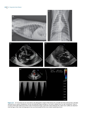
Figure 12.3 On the lateral (a) and ventrodorsal (b) radiographic images of the thorax of a 3-month-old kitten, the trachea is dorsally
elevated due to cardiac enlargement. On the ventrodorsal image, the apex of the heart is shifted into the right hemithorax. There is
rounding of the left ventricular border on the ventrodorsal image. (c,d) On the echocardiogram, the mitral valve leaflets are attached
near the apex of the heart. (e) Regurgitant flow was documented at the mitral valves measuring 6.0 m/s.