
Page 197 - Feline diagnostic imaging
P. 197
200 12 Congenital Heart Disease
(a) (b)
(c)
(d)
(e) (f)
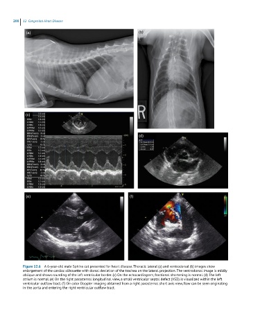
Figure 12.6 A 6-year-old male Sphinx cat presented for heart disease. Thoracic lateral (a) and ventrodorsal (b) images show
enlargement of the cardiac silhouette with dorsal deviation of the trachea on the lateral projection. The ventrodorsal image is mildly
oblique and shows rounding of the left ventricular border. (c) On the echocardiogram, fractional shortening is normal. (d) The left
atrium is normal. (e) On the right parasternal longitudinal view, a small ventricular septal defect (VSD) is visualized within the left
ventricular outflow tract. (f) On color Doppler imaging obtained from a right parasternal short axis view, flow can be seen originating
in the aorta and entering the right ventricular outflow tract.