
Page 196 - Feline diagnostic imaging
P. 196
(b)
(a)
(c) (d)
(e) (f)
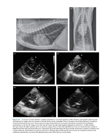
Figure 12.5 A 2.5-year-old male domestic longhair presented for increased respiratory effort. Thoracic radiographic lateral (a) and
ventrodorsal (b) images show an increase in the size of the cardiac silhouette. There is increased size of the pulmonary vasculature
including both arteries and veins. The cranial and caudal lobar arteries are enlarged and tortuous consistent with pulmonary
hypertension. (c) On the 2D image of the right and left heart, the right heart appears enlarged. LA, left atrium; LV, left ventricle;
RA, right atrium; RV, right ventricle. (d) On a transverse image of the left (LV) and right (RV) ventricles, the interventricular septum is
flattened and the right ventricular free wall is thickened. (e) At the heart base, the dorsal border of the aorta is absent, consistent with
a large ventricular septal defect. Ao, aorta. LA, left atrium. (f) Both right and left caudal lobar arteries are enlarged consistent with
pulmonary hypertension. Ao, aorta; LPA, left pulmonary artery; RPA, right pulmonary artery.