
Page 200 - Feline diagnostic imaging
P. 200
12.7 Tetralogy lof raale 203
(b)
(a)
(c) (e) (g)
(d) (f)
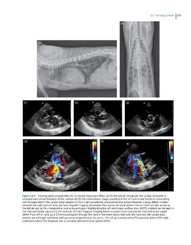
Figure 12.9 A young adult cat presented for increased respiratory effort. (a) On the lateral radiograph, the cardiac silhouette is
enlarged with dorsal deviation of the trachea. (b) On the ventrodorsal image, rounding of the left ventricular border is noted along
with enlargement of the caudal lobar arteries. (c) On a right parasternal longitudinal axis echocardiogram, a large defect is noted
between the right and left atria. (d) Color Doppler imaging documents flow across the atrial defect moving from the right atrium to
the left atrium. (e) On a longitudinal axis echocardiogram highlighting the left ventricular outflow tract (LVOT), a defect can be seen in
the interventricular septum. LV, left ventricle. (f) Color Doppler imaging shows turbulent flow crossing the interventricular septal
defect from left to right. (g) A 2D echocardiogram through the base of the heart shows that both the right and left caudal lobar
arteries are enlarged consistent with pulmonary hypertension. Ao, aorta; LPA, left pulmonary artery. PV, pulmonic artery; RPA, right
pulmonary artery. The diagnosis was a complete atrioventricular septal defect.