
Page 204 - Feline diagnostic imaging
P. 204
13.2 ccocardiograms 207
(b)
(a)
(c)
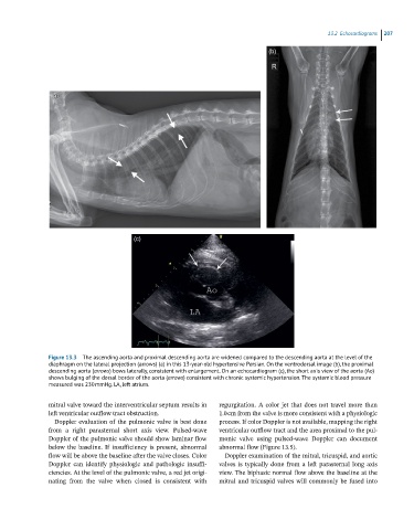
Figure 13.3 The ascending aorta and proximal descending aorta are widened compared to the descending aorta at the level of the
diaphragm on the lateral projection (arrows) (a) in this 13-year-old hypertensive Persian. On the ventrodorsal image (b), the proximal
descending aorta (arrows) bows laterally, consistent with enlargement. On an echocardiogram (c), the short axis view of the aorta (Ao)
shows bulging of the dorsal border of the aorta (arrows) consistent with chronic systemic hypertension. The systemic blood pressure
measured was 230 mmHg. LA, left atrium.
mitral valve toward the interventricular septum results in regurgitation. A color jet that does not travel more than
left ventricular outflow tract obstruction. 1.0 cm from the valve is more consistent with a physiologic
Doppler evaluation of the pulmonic valve is best done process. If color Doppler is not available, mapping the right
from a right parasternal short axis view. Pulsed‐wave ventricular outflow tract and the area proximal to the pul
Doppler of the pulmonic valve should show laminar flow monic valve using pulsed‐wave Doppler can document
below the baseline. If insufficiency is present, abnormal abnormal flow (Figure 13.5).
flow will be above the baseline after the valve closes. Color Doppler examination of the mitral, tricuspid, and aortic
Doppler can identify physiologic and pathologic insuffi valves is typically done from a left parasternal long axis
ciencies. At the level of the pulmonic valve, a red jet origi view. The biphasic normal flow above the baseline at the
nating from the valve when closed is consistent with mitral and tricuspid valves will commonly be fused into