
Page 206 - Feline diagnostic imaging
P. 206
(a)
(b)
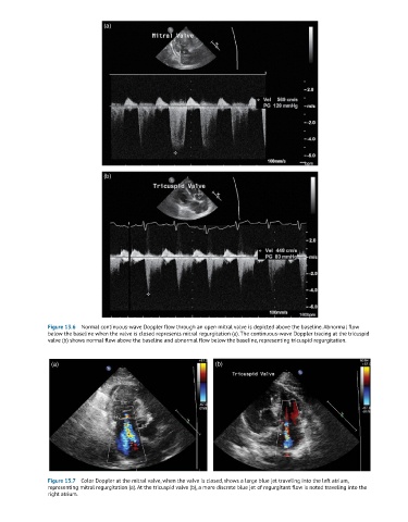
Figure 13.6 Normal continuous-wave Doppler flow through an open mitral valve is depicted above the baseline. Abnormal flow
below the baseline when the valve is closed represents mitral regurgitation (a). The continuous-wave Doppler tracing at the tricuspid
valve (b) shows normal flow above the baseline and abnormal flow below the baseline, representing tricuspid regurgitation.
(a) (b)
Figure 13.7 Color Doppler at the mitral valve, when the valve is closed, shows a large blue jet traveling into the left atrium,
representing mitral regurgitation (a). At the tricuspid valve (b), a more discrete blue jet of regurgitant flow is noted traveling into the
right atrium.