
Page 211 - Feline diagnostic imaging
P. 211
214 13 Acquired Heart Disease
(a) (b)
(c)
(d)
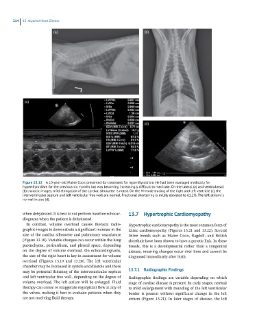
Figure 13.12 A 10-year-old Maine Coon presented for treatment for hyperthyroidism. He had been managed medically for
hyperthyroidism for the previous six months but was becoming increasingly difficult to medicate. On the lateral (a) and ventrodorsal
(b) thoracic images, mild elongation of the cardiac silhouette is noted. On the M-mode tracing of the right and left ventricle (c), the
interventricular septum and left ventricular free wall are normal. Fractional shortening is mildly elevated to 61.2%. The left atrium is
normal in size (d).
when dehydrated. It is best to not perform baseline echocar 13.7 Hypertrophic Cardiomyopathy
diograms when the patient is dehydrated.
In contrast, volume overload causes thoracic radio Hypertrophic cardiomyopathy is the most common form of
graphic images to demonstrate a significant increase in the feline cardiomyopathy (Figures 13.21 and 13.22). Several
size of the cardiac silhouette and pulmonary vasculature feline breeds such as Maine Coon, Ragdoll, and British
(Figure 13.18). Variable changes can occur within the lung shorthair have been shown to have a genetic link. In these
parenchyma, pericardium, and pleural space, depending breeds, this is a developmental rather than a congenital
on the degree of volume overload. On echocardiograms, disease, meaning changes occur over time and cannot be
the size of the right heart is key in assessment for volume diagnosed immediately after birth.
overload (Figures 13.19 and 13.20). The left ventricular
chamber may be increased in systole and diastole and there
may be potential thinning of the interventricular septum 13.7.1 Radiographic Findings
and left ventricular free wall, depending on the degree of Radiographic findings are variable depending on which
volume overload. The left atrium will be enlarged. Fluid stage of cardiac disease is present. In early stages, normal
therapy can create or exaggerate regurgitant flow at any of to mild enlargement with rounding of the left ventricular
the valves, making it best to evaluate patients when they border is present without significant change in the left
are not receiving fluid therapy. atrium (Figure 13.21). In later stages of disease, the left