
Page 210 - Feline diagnostic imaging
P. 210
13.6 Volume DepletionnVolume Oerload 213
(b)
(a)
(c)
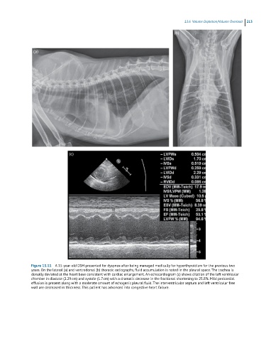
Figure 13.11 A 15-year-old DSH presented for dyspnea after being managed medically for hyperthyroidism for the previous two
years. On the lateral (a) and ventrodorsal (b) thoracic radiographs, fluid accumulation is noted in the pleural space. The trachea is
dorsally deviated at the heart base consistent with cardiac enlargement. An echocardiogram (c) shows dilation of the left ventricular
chamber in diastole (2.29 cm) and systole (1.7 cm) with a dramatic decrease in the fractional shortening to 25.8%. Mild pericardial
effusion is present along with a moderate amount of echogenic pleural fluid. The interventricular septum and left ventricular free
wall are decreased in thickness. This patient has advanced into congestive heart failure.