
Page 213 - Feline diagnostic imaging
P. 213
216 13 Acquired Heart Disease
(a) (b)
(c)
(d)
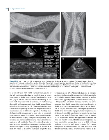
Figure 13.14 An 11-year-old DSH presented for nasal discharge. On the lateral (a) and ventrodorsal (b) thoracic images, there is
rounding of the caudal border of the cardiac silhouette. The aorta is undulating on the lateral image. On the ventrodorsal image, there
is lateral bowing of the proximal descending aorta. The echocardiogram (c) shows thickening of the interventricular septum 0.773 cm
and left ventricular free wall 0.723 cm with a normal fractional shortening of 55.7%. The aorta (arrows) has an abnormal dorsal
contour consistent with chronic systemic hypertension (d).
be commonly seen with HCM. Markedly reduced size of 7.5 mm as severe [17]. Differential diagnosis in cats pre
the left ventricular chamber in systole is due to severe senting with hypertrophic changes to the left ventricular
hypertrophy of the left ventricular walls and papillary mus wall includes systemic hypertension, acromegaly, outflow
cles (Figure 13.22). Since asymmetric thickening of the obstruction, hyperthyroidism, and volume depletion [16].
heart wall may occur with this disease, M‐mode tracing The size of the left atrium increases over time and can be
along with wall measurements from the 2D image of thick measured from the 2D image at the heart base. The ratio of
ening have been advocated [2]. Other cardiologists meas the left atrium to the aorta reportedly should be less than 1.3
ure the wall changes only from the 2D images but sequential [19] to 1.5 [20]. However, other cardiologists just measure
measurement can be difficult to reproduce. Papillary mus the left atrial size from the right parasternal longitudinal axis
cle hypertrophic changes can be seen prior to concentric 2D image. Normal left atrial size was reported as less than
hypertrophic changes. The papillary muscles will be wider 16 mm in one study [21] and less than 15.7 mm in another
at their base with varying changes in echogenicity due to [3]. In larger feline breeds, the upper level of normal left
ischemia and secondary fibrosis [17]. Measurement of the atrial size was 17–18 mm. Mild left atrial enlargement has
septum or left ventricular free wall in diastole greater or been reported in average‐sized cats to be 16.0–19.9 mm,
equal to 6 mm is considered consistent with hypertrophy. moderate 20–24 mm, and severe >24 mm [21]. Severe
Some cardiologists classify hypertrophy of 6–6.5 mm as enlargement of the left atrium has been associated with
mild, 6.6–7 mm as moderate, and equal or greater than thromboembolism. Smoke or spontaneous contrast in the