
Page 216 - Feline diagnostic imaging
P. 216
13.9 estrictiOe Cardiomyopatcy 219
(b)
(a)
(c)
(d)
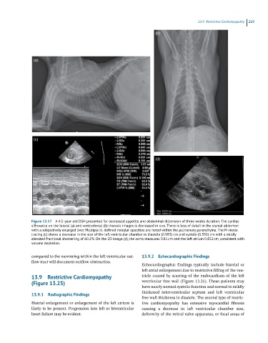
Figure 13.17 A 4.5-year-old DSH presented for decreased appetite and abdominal distension of three weeks duration. The cardiac
silhouette on the lateral (a) and ventrodorsal (b) thoracic images is decreased in size. There is loss of detail in the cranial abdomen
with a subjectively enlarged liver. Multiple ill-defined nodular opacities are noted within the pulmonary parenchyma. The M-mode
tracing (c) shows a decrease in the size of the left ventricular chamber in diastole (0.983) cm and systole (0.391) cm with a mildly
elevated fractional shortening of 60.2%. On the 2D image (d), the aorta measures 0.81 cm and the left atrium 0.852 cm, consistent with
volume depletion.
compared to the narrowing within the left ventricular out 13.9.2 Echocardiographic Findings
flow tract will document outflow obstruction.
Echocardiographic findings typically include biatrial or
left atrial enlargement due to restrictive filling of the ven
13.9 Restrictive Cardiomyopathy tricle caused by scarring of the endocardium of the left
(Figure 13.25) ventricular free wall (Figure 13.25). These patients may
have nearly normal systolic function and normal to mildly
thickened interventricular septum and left ventricular
13.9.1 Radiographic Findings
free wall thickness in diastole. The second type of restric
Biatrial enlargement or enlargement of the left atrium is tive cardiomyopathy has extensive myocardial fibrosis
likely to be present. Progression into left or biventricular causing a decrease in left ventricular chamber size,
heart failure may be evident. deformity of the mitral valve apparatus, or focal areas of