
Page 221 - Feline diagnostic imaging
P. 221
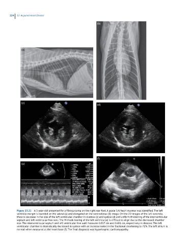
224  13  Acquired Heart Disease

                                                               (b)


               (a)


               (c)
                                                               (d)


                                                               (f)

               (e)


            Figure 13.21  A 5-year-old presented for a fibrosarcoma on the right rear foot. A grade 5/6 heart murmur was identified. The left
            ventricle margin is rounded on the lateral (a) and elongated on the ventrodorsal (b) image. On the 2D images of the left ventricle,
            there is decrease in the size of the left ventricular chamber in diastole (c) and systole (d) and uniform thickening of the interventricular
            septum and left ventricular free wall. The M-mode tracing of the left ventricle (e) is difficult to align due to the decreased chamber
            size. The interventricular septum and left ventricular free wall measures 0.857 cm and 0.681 cm, respectively in diastole. The left
            ventricular chamber is dramatically decreased in systole with an increase noted in the fractional shortening to 71%. The left atrium is
            normal when measured at the heart base (f). The final diagnosis was hypertrophic cardiomyopathy.