
Page 222 - Feline diagnostic imaging
P. 222
13.12 Arrcytcmogenic igct Ventricular Cardiomyopatcy 225
(a) (b)
(c)
(d) (e)
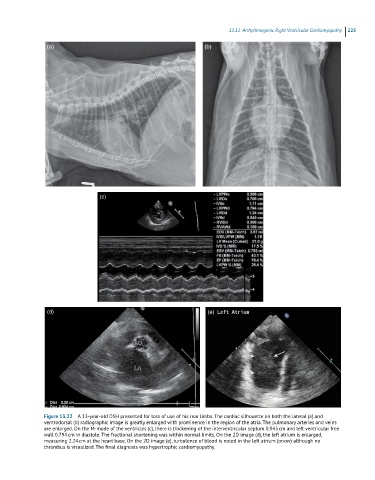
Figure 13.22 A 13-year-old DSH presented for loss of use of his rear limbs. The cardiac silhouette on both the lateral (a) and
ventrodorsal (b) radiographic image is greatly enlarged with prominence in the region of the atria. The pulmonary arteries and veins
are enlarged. On the M-mode of the ventricles (c), there is thickening of the interventricular septum 0.945 cm and left ventricular free
wall 0.794 cm in diastole. The fractional shortening was within normal limits. On the 2D image (d), the left atrium is enlarged,
measuring 2.24 cm at the heart base. On the 2D image (e), turbulence of blood is noted in the left atrium (arrow) although no
thrombus is visualized. The final diagnosis was hypertrophic cardiomyopathy.