
Page 227 - Feline diagnostic imaging
P. 227
230 13 Acquired Heart Disease
(b)
(a)
(c) (d)
(e)
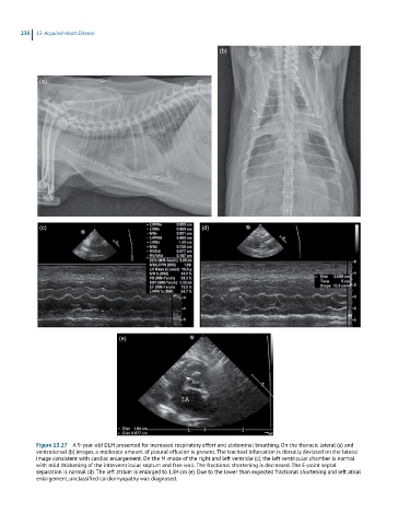
Figure 13.27 A 9-year-old DLH presented for increased respiratory effort and abdominal breathing. On the thoracic lateral (a) and
ventrodorsal (b) images, a moderate amount of pleural effusion is present. The tracheal bifurcation is dorsally deviated on the lateral
image consistent with cardiac enlargement. On the M-mode of the right and left ventricle (c), the left ventricular chamber is normal
with mild thickening of the interventricular septum and free wall. The fractional shortening is decreased. The E-point septal
separation is normal (d). The left atrium is enlarged to 1.84 cm (e). Due to the lower than expected fractional shortening and left atrial
enlargement, unclassified cardiomyopathy was diagnosed.