
Page 231 - Feline diagnostic imaging
P. 231
234 13 Acquired Heart Disease
(a) (c)
(b) (d) (e)
(f) (g)
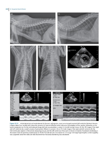
Figure 13.31 Initial lateral (a) and ventrodorsal (b) thoracic radiographs reveal an enlarged rounded left ventricle. Recheck thoracic
images, taken four months later, show the presence of pleural effusion which obscures visualization of the cardiac silhouette on the
lateral projection (c). On the ventrodorsal image (d), fluid accumulation is noted in the left cranial thorax. On the 2D image of the right
and left ventricle (e), a small volume of pericardial effusion is present. On an M-mode image of the right and left ventricle (f), the
interventricular septum is thickened, measuring 0.6 cm, and the left ventricular free wall is 0.8 cm in diastole. Fractional shortening is
decreased from the previous examination to 33.0%. The left atrium is increased to 2.2 cm (g). End-stage hypertrophic cardiomyopathy
was suspected since the walls are still thickened but fractional shortening has decreased.