
Page 232 - Feline diagnostic imaging
P. 232
13.14 Arteriotcromboembolism 235
(a) (b)
(c)
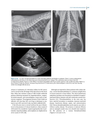
Figure 13.32 A 4-year-old DLH presented for acute respiratory distress and hindlimb weakness. There is cardiac enlargement
predominantly left-sided on the lateral (a) and ventrodorsal (b) thoracic images. The pulmonary veins are larger than the
corresponding arteries. There is a mild diffuse interstitial lung pattern with focal alveolar opacity in the caudal lung lobes. There is a
small volume of pleural effusion. Increased lung interference is noted during the echocardiogram. A thrombus was seen moving
within the enlarged left atrium (c).
atrium or localization of a thrombus within the left auricle Although not reported in feline patients with cardiac dis
can be viewed on the 2D image of the left atrium at the heart ease, venous thromboembolism is a common complication
base. Other less common causes of ATE include euthyroid in human patients in heart failure. The risk of pulmonary
patients following treatment for hyperthyroidism, congeni embolism and deep vein thrombosis is doubled in hospital
tal mitral stenosis, and noncardiogenic causes such as pul ized heart failure patients with left ventricular systolic dys
monary neoplasia. The prognosis worsens if both limbs are function [31]. Thromboembolism in the vena cava has
affected, with less than 40% surviving to discharge in con been reported secondary to neoplasia, immune‐mediated
trast to up to 80% survival if only one limb is affected [29]. disease, heartworm disease, sepsis, and catheterization
If the left atrial size is normal (without the use of diuret [32]. Venous thromboembolism can be seen in cats with or
ics or in the presence of volume depletion), other causes of without cardiac disease. The presence of pleural effusion
ATE need to be considered. Hypercoagulopathy due to pro and an enlarged caudal vena cava should raise suspicion
teinuria, hyperthyroidism, and neoplasia are implicated as for the presence of a venous thromboembolism
other causes of ATE [30]. (Figures 13.36 and 13.37).