
Page 228 - Feline diagnostic imaging
P. 228
13.14 Arteriotcromboembolism 231
(a) (b)
(c)
(d)
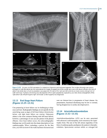
Figure 13.28 A 6-year-old DLH presented for abdominal distension and decreased appetite. The cardiac silhouette was greatly
enlarged on both the lateral (a) and ventrodorsal (b) images. Enlarged size of the caudal vena cava with pleural effusion and loss of
abdominal detail due to fluid accumulation was apparent. M-mode of the right and left ventricle (c) shows severe enlargement of
both ventricles. Fractional shortening was decreased to 13%. A right parasternal longitudinal axis view shows enlargement of the
right atrium (d). Arrhythmogenic right ventricular cardiomyopathy was diagnosed.
13.13 End-Stage Heart Failure now are thinned due to progression of heart disease. On
(Figures 13.29–13.31) presentation, fractional shortening may be low or normal,
having dropped due to cardiac decompensation.
Cats presenting in heart failure can be challenging to diag
nose and treat. Radiographic findings are not specific for the
underlying cause as signs of left heart failure, biventricular 13.14 Arteriothromboembolism
failure, and right heart failure can overlap. Pulmonary (Figures 13.32–13.35)
edema is the most common finding with left heart failure;
however, a percentage of cats can also present with pleural Arteriothromboembolism (ATE) can be seen associated
effusion. Once heart failure occurs, the echocardiographic with advanced cardiomyopathy, most commonly the hyper
findings may not fit neatly into the classic description of one trophic form. The size of the left atrium regardless of the
cardiomyopathy. When evaluating a patient for the first form of cardiomyopathy predisposes the patient to thrombo
time, you will not know if the walls had been thickened and embolism. Spontaneous contrast within an enlarged left