
Page 226 - Feline diagnostic imaging
P. 226
13.12 Arrcytcmogenic igct Ventricular Cardiomyopatcy 229
(a) (b)
(a)
(b)
(c)
(c)
(e)
(d) (e)
(d)
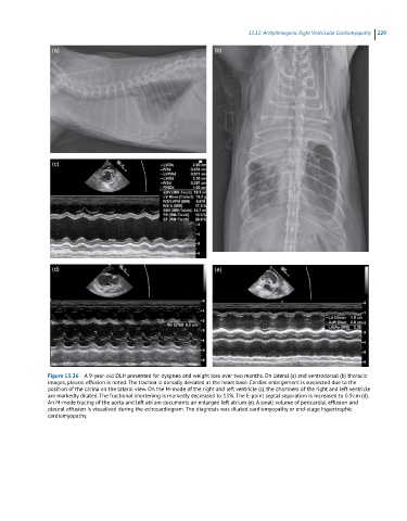
Figure 13.26 A 9-year-old DLH presented for dyspnea and weight loss over two months. On lateral (a) and ventrodorsal (b) thoracic
images, pleural effusion is noted. The trachea is dorsally deviated at the heart base. Cardiac enlargement is suspected due to the
position of the carina on the lateral view. On the M-mode of the right and left ventricle (c), the chambers of the right and left ventricle
are markedly dilated. The fractional shortening is markedly decreased to 13%. The E-point septal separation is increased to 0.9 cm (d).
An M-mode tracing of the aorta and left atrium documents an enlarged left atrium (e). A small volume of pericardial effusion and
pleural effusion is visualized during the echocardiogram. The diagnosis was dilated cardiomyopathy or end-stage hypertrophic
cardiomyopathy.