
Page 224 - Feline diagnostic imaging
P. 224
13.12 Arrcytcmogenic igct Ventricular Cardiomyopatcy 227
(a) (b)
(c)
(d)
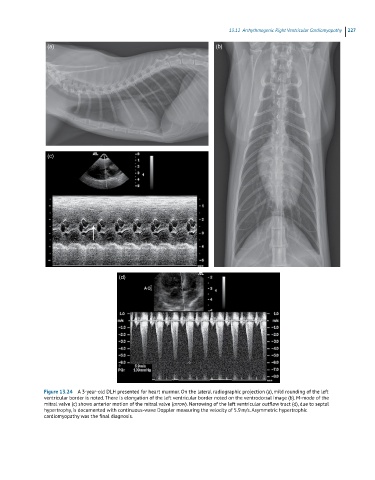
Figure 13.24 A 3-year-old DLH presented for heart murmur. On the lateral radiographic projection (a), mild rounding of the left
ventricular border is noted. There is elongation of the left ventricular border noted on the ventrodorsal image (b). M-mode of the
mitral valve (c) shows anterior motion of the mitral valve (arrow). Narrowing of the left ventricular outflow tract (d), due to septal
hypertrophy, is documented with continuous-wave Doppler measuring the velocity of 5.9 m/s. Asymmetric hypertrophic
cardiomyopathy was the final diagnosis.