
Page 229 - Feline diagnostic imaging
P. 229
232 13 Acquired Heart Disease
(a) (b)
(c)
(d)
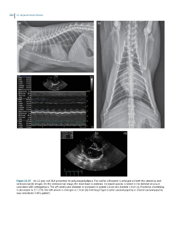
Figure 13.29 An 11-year-old DLH presented for polyuria/polydipsia. The cardiac silhouette is enlarged on both the lateral (a) and
ventrodorsal (b) images. On the ventrodorsal image, the heart base is widened. Increased opacity is noted in the skeletal structure
consistent with osteopetrosis. The left ventricular chamber is increased in systole 2.0 cm and diastole 1.4 cm (c). Fractional shortening
is decreased to 21–27%. The left atrium is enlarged at 1.9 cm (d). End-stage hypertrophic cardiomyopathy or dilated cardiomyopathy
was considered in this patient.