
Page 233 - Feline diagnostic imaging
P. 233
236 13 Acquired Heart Disease
(a) (b)
(c)
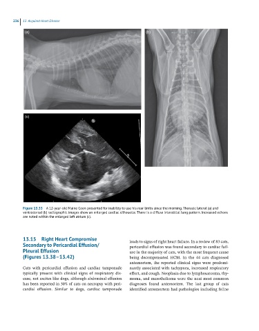
Figure 13.33 A 12-year-old Maine Coon presented for inability to use his rear limbs since the morning. Thoracic lateral (a) and
ventrodorsal (b) radiographic images show an enlarged cardiac silhouette. There is a diffuse interstitial lung pattern. Increased echoes
are noted within the enlarged left atrium (c).
13.15 Right Heart Compromise leads to signs of right heart failure. In a review of 83 cats,
Secondary to Pericardial Effusion/ pericardial effusion was found secondary to cardiac fail
Pleural Effusion ure in the majority of cats, with the most frequent cause
(Figures 13.38–13.42) being decompensated HCM. In the 44 cats diagnosed
antemortem, the reported clinical signs were predomi
Cats with pericardial effusion and cardiac tamponade nantly associated with tachypnea, increased respiratory
typically present with clinical signs of respiratory dis effort, and cough. Neoplasia due to lymphosarcoma, thy
ease, not ascites like dogs, although abdominal effusion moma, and mesothelioma were the next most common
has been reported in 30% of cats on necropsy with peri diagnoses found antemortem. The last group of cats
cardial effusion. Similar to dogs, cardiac tamponade identified antemortem had pathologies including feline