
Page 238 - Feline diagnostic imaging
P. 238
13.17 eoplasia 241
(a) (b)
(c)
(d)
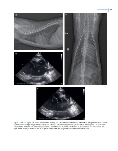
Figure 13.38 An 8-year-old Persian presented for fainting and a heart murmur. The cardiac silhouette is enlarged on both the lateral
(a) and ventrodorsal (b) images. A heavy interstitial pattern is present most significantly in the left caudal lung lobe. The pulmonary
vasculature is enlarged. The echocardiogram reveals a small volume of pericardial effusion (c). Mild collapse and flattening of the
right atrium (arrows) is noted on the 2D image (d). This patient was diagnosed with congestive heart failure.