
Page 239 - Feline diagnostic imaging
P. 239
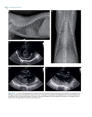
242  13  Acquired Heart Disease

             (a)                                                      (b)


             (c)


             (d)                                              (e)


            Figure 13.39  A 5-year-old DSH presented for diabetes and a heart murmur. Thoracic lateral (a) and ventrodorsal (b) images show an
            enlarged rounded cardiac silhouette. The caudal vena cava is enlarged as it approaches the diaphragm best seen on the lateral view.
            A moderate amount of pericardial effusion is present on the echocardiogram (c). Mild collapse of the free wall of the right atrium is
            noted on the 2D image (d,e). LA, left atrium; RA, right atrium.