
Page 242 - Feline diagnostic imaging
P. 242
13.18 Heartworm Disease 245
(a) (b)
(c) (d)
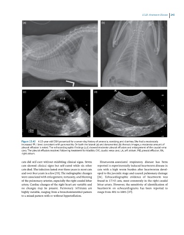
Figure 13.42 A 13-year-old DSH presented for a seven-day history of anorexia, vomiting, and diarrhea. She had a moderately
increased fPLI level consistent with pancreatitis. On both the lateral (a) and dorsoventral (b) thoracic images, a moderate amount of
pleural effusion is noted. The echocardiographic findings (c,d) showed moderate pleural effusion and enlargement of the caudal vena
cava. The pleural effusion resolved following treatment for triaditis. CVC, caudal vena cava; LA, left atrium; PlE, pleural effusion; RA,
right atrium.
cats did self‐cure without exhibiting clinical signs. Seven Heartworm‐associated respiratory disease has been
cats showed clinical signs but self‐cured while six other reported in experimentally induced heartworm disease in
cats died. The infection lasted over three years in most cats cats with a high worm burden after heartworms devel
and over four years in a few [35]. The radiographic changes oped to the juvenile stage and caused pulmonary damage
were associated with enlargement, tortuosity, and blunting [36]. Echocardiographic evidence of heartworm was
of the pulmonary arteries, especially the right caudal lobar found in 17/43 cats, most commonly in the right caudal
artery. Cardiac changes of the right heart are variable and lobar artery. However, the sensitivity of identification of
no changes may be present. Pulmonary infiltrates are heartworm on echocardiograms has been reported to
highly variable, ranging from a bronchointerstitial pattern range from 40% to 100% [37].
to a mixed pattern with or without hyperinflation.