
Page 244 - Feline diagnostic imaging
P. 244
13.19 igct Ventricular utflow bstruction 247
(b)
(a)
(c) (d)
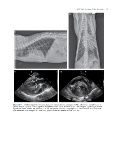
Figure 13.44 Both lateral (a) and ventrodorsal (b) thoracic radiographs show enlargement of the left ventricle. A small volume of
pleural effusion is present. There is slightly decreased detail in the cranial abdomen. On the echocardiogram (c), a focally thickened
hyperechoic area is noted in the right ventricular free wall (arrows). On the 2D image (d), the left ventricular wall is thickened with
mild alterations in the echogenicity. On necropsy, lymphosarcoma was found within the heart wall.