
Page 240 - Feline diagnostic imaging
P. 240
13.18 Heartworm Disease 243
(a) (b)
(c)
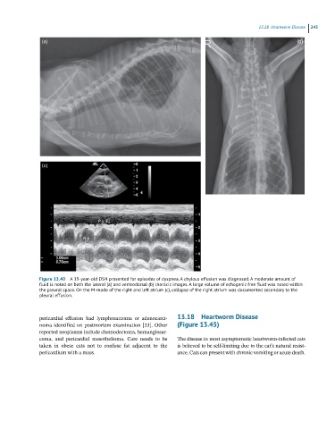
Figure 13.40 A 19-year-old DSH presented for episodes of dyspnea. A chylous effusion was diagnosed. A moderate amount of
fluid is noted on both the lateral (a) and ventrodorsal (b) thoracic images. A large volume of echogenic free fluid was noted within
the pleural space. On the M-mode of the right and left atrium (c), collapse of the right atrium was documented secondary to the
pleural effusion.
pericardial effusion had lymphosarcoma or adenocarci 13.18 Heartworm Disease
noma identified on postmortem examination [33]. Other (Figure 13.45)
reported neoplasms include chemodectoma, hemangiosar
coma, and pericardial mesothelioma. Care needs to be The disease in most asymptomatic heartworm‐infected cats
taken in obese cats not to confuse fat adjacent to the is believed to be self‐limiting due to the cat’s natural resist
pericardium with a mass. ance. Cats can present with chronic vomiting or acute death.