
Page 237 - Feline diagnostic imaging
P. 237
240 13 Acquired Heart Disease
(a) (b)
(c)
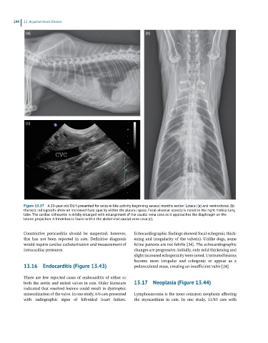
Figure 13.37 A 20-year-old DLH presented for seizure-like activity beginning several months earlier. Lateral (a) and ventrodorsal (b)
thoracic radiographs show an increased fluid opacity within the pleural space. Focal alveolar opacity is noted in the right middle lung
lobe. The cardiac silhouette is mildly enlarged with enlargement of the caudal vena cava as it approaches the diaphragm on the
lateral projection. A thrombus is found within the abdominal caudal vena cava (c).
Constrictive pericarditis should be suspected; however, Echocardiographic findings showed focal echogenic thick
this has not been reported in cats. Definitive diagnosis ening and irregularity of the valve(s). Unlike dogs, some
would require cardiac catheterization and measurement of feline patients are not febrile [34]. The echocardiographic
intracardiac pressures. changes are progressive; initially, only mild thickening and
slight increased echogenicity were noted. Untreated lesions
become more irregular and echogenic or appear as a
13.16 Endocarditis (Figure 13.43) pedunculated mass, creating an insufficient valve [24].
There are few reported cases of endocarditis of either or
both the aortic and mitral valves in cats. Older literature 13.17 Neoplasia (Figure 13.44)
indicated that resolved lesions could result in dystrophic
mineralization of the valve. In one study, 4/6 cats presented Lymphosarcoma is the most common neoplasm affecting
with radiographic signs of left‐sided heart failure. the myocardium in cats. In one study, 11/83 cats with