
Page 236 - Feline diagnostic imaging
P. 236
(a) (b)
(c) (d)
(e)
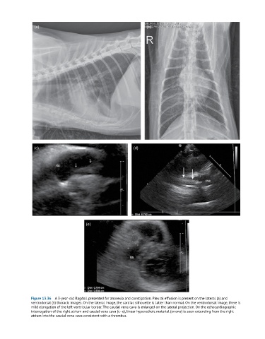
Figure 13.36 A 3-year-old Ragdoll presented for anorexia and constipation. Pleural effusion is present on the lateral (a) and
ventrodorsal (b) thoracic images. On the lateral image, the cardiac silhouette is taller than normal. On the ventrodorsal image, there is
mild elongation of the left ventricular border. The caudal vena cava is enlarged on the lateral projection. On the echocardiographic
interrogation of the right atrium and caudal vena cava (c–e), linear hyperechoic material (arrows) is seen extending from the right
atrium into the caudal vena cava consistent with a thrombus.