
Page 241 - Feline diagnostic imaging
P. 241
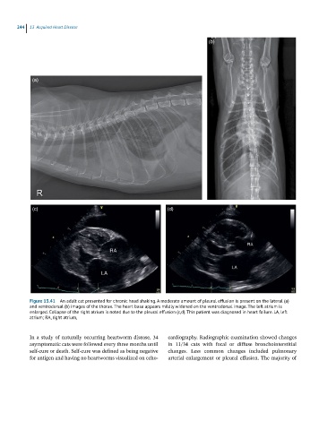
244 13 Acquired Heart Disease
(b)
(a)
(c) (d)
Figure 13.41 An adult cat presented for chronic head shaking. A moderate amount of pleural effusion is present on the lateral (a)
and ventrodorsal (b) images of the thorax. The heart base appears mildly widened on the ventrodorsal image. The left atrium is
enlarged. Collapse of the right atrium is noted due to the pleural effusion (c,d). This patient was diagnosed in heart failure. LA, left
atrium; RA, right atrium,
In a study of naturally occurring heartworm disease, 34 cardiography. Radiographic examination showed changes
asymptomatic cats were followed every three months until in 11/34 cats with focal or diffuse bronchointerstitial
self‐cure or death. Self‐cure was defined as being negative changes. Less common changes included pulmonary
for antigen and having no heartworms visualized on echo arterial enlargement or pleural effusion. The majority of