
Page 245 - Feline diagnostic imaging
P. 245
248 13 Acquired Heart Disease
(b)
(a)
(c) (d)
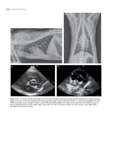
Figure 13.45 A 16-year-old DSH presented for an oral squamous cell carcinoma involving the left mandible. The cardiac silhouette
appears enlarged on both the lateral (a) and ventrodorsal (b) images. The left ventricular border is rounded and elongated. The right
middle lung lobe appears collapsed. There is a mild diffuse bronchial pattern. The right caudal lobar artery was mildly enlarged. A
right parasternal short axis view reveals “equal” signs within the right ventricular outflow tract and the right caudal lobar artery
consistent with heartworms (c,d).