
Page 246 - Feline diagnostic imaging
P. 246
13.19 igct Ventricular utflow bstruction 249
(b)
(a)
(c) (d)
(e)
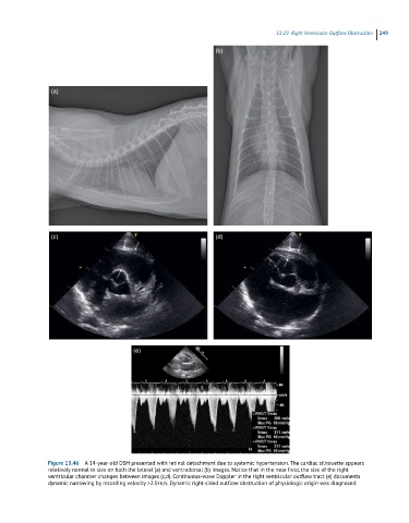
Figure 13.46 A 14-year-old DSH presented with retinal detachment due to systemic hypertension. The cardiac silhouette appears
relatively normal in size on both the lateral (a) and ventrodorsal (b) images. Notice that in the near field, the size of the right
ventricular chamber changes between images (c,d). Continuous-wave Doppler in the right ventricular outflow tract (e) documents
dynamic narrowing by recording velocity >2.0 m/s. Dynamic right-sided outflow obstruction of physiologic origin was diagnosed.