
Page 251 - Feline diagnostic imaging
P. 251
14.1 Paaterns of DnstPnst 255
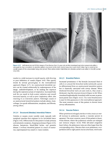
(b)
(a)
Figure 14.3 Left lateral (a) and VD (b) images of the thorax of an 11-year-old cat that developed aspiration pneumonia after a
laryngeal tie-back procedure. An alveolar pattern is present in the right middle lung lobe. A prominent lobar sign is noted at the
caudal aspect of the consolidated lung lobe (arrow). More subtle mixed interstitial and alveolar patterns are noted in the left cranial
lung lobe.
results in a mild increase in overall opacity, with blurring 14.1.5 Bronchial Pattern
or poor definition of vessels (Figure 14.6). This opacity Increased prominence of the bronchi (increased fluid or
would be termed ground‐glass in the nontraditional
approach (Figure 14.7). An unstructured interstitial pat- cells in or around the bronchial walls) technically cannot
be differentiated from an unstructured interstitial pattern
tern can be created artifactually by underexposure of the
image, underdevelopment, or by making the exposure but is classically associated with airway disease. When
thickened bronchial walls are seen end‐on, they create a
during the expiratory phase of respiration. Increased body
wall fat can result in both scatter radiation and overall thickened, ring‐like structure (donut) (Figure 14.10). When
visualized longitudinally, bronchial walls are seen as prom-
increased opacity, as well as poor inspiratory effort, also
resulting in an interstitial pattern. Pathologic causes of inent paired lines (Figure 14.11). The term bronchocentric
an unstructured interstitial pattern include edema, hem- is used for this appearance in the nontraditional approach.
orrhage, low‐grade inflammation, neoplasia, and fibrosis, The most common cause of this pattern is chronic lower
among others. airway inflammation.
14.1.6 Vascular Pattern
14.1.4 Structured (Nodular) Interstitial Pattern
When prominent/enlarged arteries or veins create an over-
Nodules or masses create variably sized, typically well‐ all increase in pulmonary opacity, a vascular pattern is
margined opacities that originate in the interstitial tissue apparent. The most common cause of this pattern is heart-
(Figure 14.8). Differentials for this pattern include primary worm disease, where the pulmonary arteries are enlarged
and metastatic neoplasia, fungal granulomas (Figure 14.9), and tortuous (Figure 14.12). With left‐sided congestive
abscess, hematoma, blood‐filled bullae, and parasitic heart failure, the pulmonary veins are often larger than the
disease. A miliary interstitial pattern is a result of numer- corresponding artery. Diseases that cause pulmonary over-
ous, superimposed very small (1–3 mm) nodules. perfusion (left to right patent ductus arteriosus, ventricular