
Page 256 - Feline diagnostic imaging
P. 256
260 14 Feline Pulmonary Disease
(b)
(a)
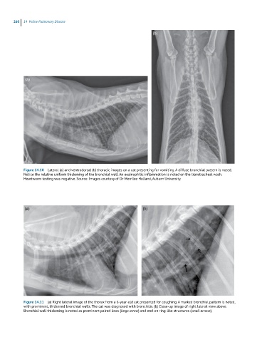
Figure 14.10 Lateral (a) and ventrodorsal (b) thoracic images on a cat presenting for vomiting. A diffuse bronchial pattern is noted.
Notice the relative uniform thickening of the bronchial wall. An eosinophilic inflammation is noted on the transtracheal wash.
Heartworm testing was negative. Source: Images courtesy of Dr Merrilee Holland, Auburn University.
(a) (b)
Figure 14.11 (a) Right lateral image of the thorax from a 6-year-old cat presented for coughing. A marked bronchial pattern is noted,
with prominent, thickened bronchial walls. The cat was diagnosed with bronchitis. (b) Close-up image of right lateral view above.
Bronchial wall thickening is noted as prominent paired lines (large arrow) and end-on ring-like structures (small arrows).