
Page 257 - Feline diagnostic imaging
P. 257
14.3 Pulmonary eoplasia 261
(b)
(a)
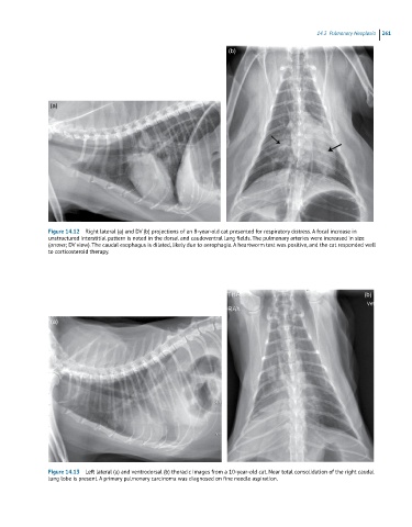
Figure 14.12 Right lateral (a) and DV (b) projections of an 8-year-old cat presented for respiratory distress. A focal increase in
unstructured interstitial pattern is noted in the dorsal and caudoventral lung fields. The pulmonary arteries were increased in size
(arrows; DV view). The caudal esophagus is dilated, likely due to aerophagia. A heartworm test was positive, and the cat responded well
to corticosteroid therapy.
(b)
(a)
Figure 14.13 Left lateral (a) and ventrodorsal (b) thoracic images from a 10-year-old cat. Near total consolidation of the right caudal
lung lobe is present. A primary pulmonary carcinoma was diagnosed on fine needle aspiration.