
Page 262 - Feline diagnostic imaging
P. 262
266 14 Feline Pulmonary Disease
(a) (b)
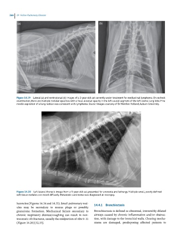
Figure 14.19 Lateral (a) and ventrodorsal (b) images of a 2-year-old cat currently under treatment for mediastinal lymphoma. On recheck
examination, there are multiple nodular opacities with a focal alveolar opacity in the left caudal segment of the left cranial lung lobe. Fine
needle aspiration of a lung nodule was consistent with lymphoma. Source: Images courtesy of Dr Merrilee Holland, Auburn University.
Figure 14.20 Left lateral thoracic image from a 9-year-old cat presented for anorexia and lethargy. Multiple small, poorly defined
soft tissue nodules are noted diffusely. Metastatic carcinoma was diagnosed at necropsy.
lucencies (Figures 14.24 and 14.25). Small pulmonary nod- 14.4.1 Bronchiectasis
ules may be secondary to mucus plugs or possibly
granuloma formation. Mechanical failure secondary to Bronchiectasis is defined as abnormal, irreversibly dilated
chronic respiratory distress/coughing can result in non- airways caused by chronic inflammation and/or obstruc-
traumatic rib fractures, usually the midportion of ribs 9–11 tion, with damage to the bronchial walls. Clearing mecha-
(Figure 14.26) [32,33]. nisms are damaged, predisposing affected patients to