
Page 266 - Feline diagnostic imaging
P. 266
270 14 Feline Pulmonary Disease
(a) (b)
(c)
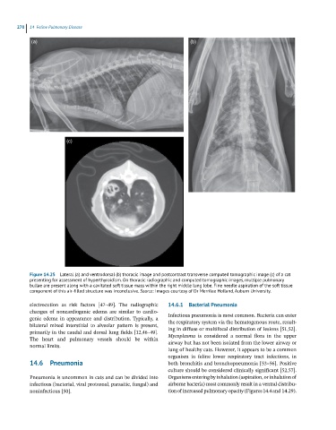
Figure 14.25 Lateral (a) and ventrodorsal (b) thoracic image and postcontrast transverse computed tomographic image (c) of a cat
presenting for assessment of hyperthyroidism. On thoracic radiographic and computed tomographic images, multiple pulmonary
bullae are present along with a cavitated soft tissue mass within the right middle lung lobe. Fine needle aspiration of the soft tissue
component of this air-filled structure was inconclusive. Source: Images courtesy of Dr Merrilee Holland, Auburn University.
electrocution as risk factors [47–49]. The radiographic 14.6.1 Bacterial Pneumonia
changes of noncardiogenic edema are similar to cardio- Infectious pneumonia is most common. Bacteria can enter
genic edema in appearance and distribution. Typically, a
bilateral mixed interstitial to alveolar pattern is present, the respiratory system via the hematogenous route, result-
ing in diffuse or multifocal distribution of lesions [51,52].
primarily in the caudal and dorsal lung fields [12,46–49].
The heart and pulmonary vessels should be within Mycoplasma is considered a normal flora in the upper
airway but has not been isolated from the lower airway or
normal limits.
lung of healthy cats. However, it appears to be a common
organism in feline lower respiratory tract infections, in
14.6 Pneumonia both bronchitis and bronchopneumonia [53–56]. Positive
culture should be considered clinically significant [52,57].
Pneumonia is uncommon in cats and can be divided into Organisms entering by inhalation (aspiration, or inhalation of
infectious (bacterial, viral protozoal, parasitic, fungal) and airborne bacteria) most commonly result in a ventral distribu-
noninfectious [50]. tion of increased pulmonary opacity (Figures 14.4 and 14.29).