
Page 267 - Feline diagnostic imaging
P. 267
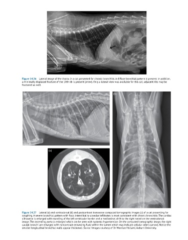
Figure 14.26  Lateral image of the thorax in a cat presented for chronic bronchitis. A diffuse bronchial pattern is present. In addition,
               a minimally displaced fracture of the 10th rib is present (arrow). Only a lateral view was available for this cat; adjacent ribs may be
               fractured as well.


                (a)                                                        (b)


                                 (c)


               Figure 14.27  Lateral (a) and ventrodorsal (b) and postcontrast transverse computed tomographic images (c) of a cat presenting for
               coughing. A severe bronchial pattern with focal interstitial to alveolar infiltrates is most consistent with chronic bronchitis. The cardiac
               silhouette is enlarged with rounding of the left ventricular border and a mediastinal shift to the right noted on the ventrodorsal
               image. The ascending aorta is enlarged which can be seen with systemic hypertension. On the computed tomographic image, the right
               caudal bronchi are enlarged with noncontrast-enhancing fluid within the lumen which may indicate cellular debris (arrow). Notice the
               smaller longitudinal bronchial walls appear thickened. Source: Images courtesy of Dr Merrilee Holland, Auburn University.