
Page 270 - Feline diagnostic imaging
P. 270
274 14 Feline Pulmonary Disease
Diffuse unstructured interstitial and alveolar patterns may related to the worm burden, and young, debilitated, or
also occur. immunosuppressed cats are more likely to have clinically
significant infections.
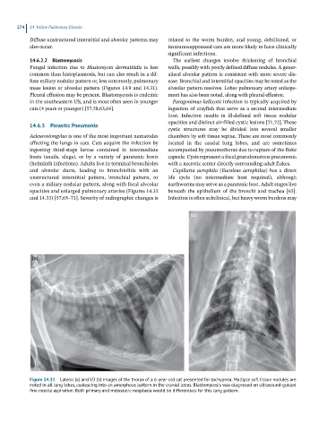
14.6.2.2 Blastomycosis The earliest changes involve thickening of bronchial
Fungal infection due to Blastomyces dermatitidis is less walls, possibly with poorly defined diffuse nodules. A gener-
common than histoplasmosis, but can also result in a dif- alized alveolar pattern is consistent with more severe dis-
fuse miliary nodular pattern or, less commonly, pulmonary ease. Bronchial and interstitial opacities may be noted as the
mass lesion or alveolar pattern (Figures 14.9 and 14.31). alveolar pattern resolves. Lobar pulmonary artery enlarge-
Pleural effusion may be present. Blastomycosis is endemic ment has also been noted, along with pleural effusion.
in the southeastern US, and is most often seen in younger Paragonimus kellicotti infection is typically acquired by
cats (4 years or younger) [57,58,63,64]. ingestion of crayfish that serve as a second intermediate
host. Infection results in ill‐defined soft tissue nodular
opacities and distinct air‐filled cystic lesions [71,72]. These
14.6.3 Parasitic Pneumonia
cystic structures may be divided into several smaller
Aeleurostongylus is one of the most important nematodes chambers by soft tissue septae. These are most commonly
affecting the lungs in cats. Cats acquire the infection by located in the caudal lung lobes, and are sometimes
ingesting third‐stage larvae contained in intermediate accompanied by pneumothorax due to rupture of the fluke
hosts (snails, slugs), or by a variety of paratenic hosts capsule. Cysts represent a focal granulomatous pneumonia
(helminth infections). Adults live in terminal bronchioles with a necrotic center directly surrounding adult flukes.
and alveolar ducts, leading to bronchiolitis with an Capillaria aerophila ( Eucoleus aerophilus) has a direct
unstructured interstitial pattern, bronchial pattern, or life cycle (no intermediate host required), although
even a miliary nodular pattern, along with focal alveolar earthworms may serve as a paratenic host. Adult stages live
opacities and enlarged pulmonary arteries (Figures 14.32 beneath the epithelium of the bronchi and trachea [65].
and 14.33) [57,65–71]. Severity of radiographic changes is Infection is often subclinical, but heavy worm burdens may
(b)
(a)
Figure 14.31 Lateral (a) and VD (b) images of the thorax of a 6-year-old cat presented for tachypnea. Multiple soft tissue nodules are
noted in all lung lobes, coalescing into an amorphous pattern in the cranial lobes. Blastomycosis was diagnosed on ultrasound-guided
fine needle aspiration. Both primary and metastatic neoplasia would be differentials for this lung pattern.