
Page 265 - Feline diagnostic imaging
P. 265
14.5 Pulmonary dema 269
(b)
(a)
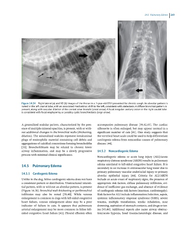
Figure 14.24 Right lateral (a) and VD (b) images of the thorax in a 7-year-old DSH presented for chronic cough. An alveolar pattern is
noted in the left cranial lobe, with an associated mediastinal shift to the left, consistent with atelectasis. A diffuse bronchial pattern is
present, along with saccular dilation of the cranial lobar bronchi (small arrow). A focal irregular cavitary lesion in the right caudal lobe
is consistent with focal emphysema, or possibly cystic bronchiectasis (large arrow).
A generalized nodular pattern, characterized by the pres- accompanies pulmonary disease [39,42,43]. The cardiac
ence of multiple mineral opacities, is present, with or with- silhouette is often enlarged, but may appear normal in a
out additional changes in the bronchial walls (thickening, significant number of cats [41]. One study suggests that
dilation). The mineralized nodules represent intraluminal the vertebral heart scale could be used to help differentiate
plugs of eosinophilic material containing cell debris and cardiogenic edema from noncardiac causes of pulmonary
aggregations of calcified concretions forming broncholiths disease [44].
[35]. Broncholithiasis may be related to chronic lower
airway inflammation, and may be a slowly progressive 14.5.2 Noncardiogenic Edema
process with minimal clinical significance.
Noncardiogenic edema or acute lung injury (ALI)/acute
respiratory distress syndrome (ARDS) results in pulmonary
14.5 Pulmonary Edema edema unrelated to left‐sided congestive heart failure. It is
secondary to an increase in extravascular lung water due to
primary pulmonary vascular endothelial injury or primary
14.5.1 Cardiogenic Edema
alveolar epithelial injury [44]. Criteria for ALI/ARDS
Unlike in the dog, feline cardiogenic edema does not have include an acute onset of respiratory signs, the presence of
a consistent pattern or distribution. Unstructured intersti- appropriate risk factors, diffuse pulmonary infiltrates, evi-
tial pattern, with or without an alveolar pattern, is present dence of inefficient gas exchange, and absence of evidence
(Figure 14.28). Bronchial wall thickening or peribronchial of cardiogenic edema risk factors (murmur, cardiomegaly).
infiltrates may also be noted [39,40]. While venous Risk factors for ALI include inflammation/infection, sepsis,
enlargement is common in dogs with left‐sided congestive systemic inflammatory response syndrome (SIRS), severe
heart failure, venous enlargement alone may be a poor trauma, multiple transfusions, smoke inhalation, near
indicator of failure in cats. It appears that pulmonary drowning, aspiration of stomach contents, and drugs or tox-
arterial enlargement may be more common in feline left‐ ins [45,46]. Additional reports also name airway obstruc-
sided congestive heart failure [41]. Pleural effusion often tion/acute hypoxia, head trauma/neurologic disease, and