
Page 260 - Feline diagnostic imaging
P. 260
264 14 Feline Pulmonary Disease
(a) (b)
(c)
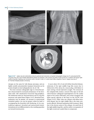
Figure 14.17 Lateral (a) and ventrodorsal (b) and postcontrast transverse computed tomographic image (c) of a cat presented for
coughing. Two soft tissue focally mineralized masses are present in the caudal lung lobes. Focal cavitation is noted in the larger right
caudal lung lobe. Cytology was consistent with a necrotic center of a mass, most likely neoplasia. Source: Images courtesy of
Dr Merrilee Holland, Auburn University.
changes are the same for both disease processes, and are In up to about 10% of cats with feline bronchial disease,
highly variable and potentially transient; bronchitis can be atelectasis of the right middle lung lobe occurs due to
present despite normal radiographs [22–24, 26–30]. obstruction of the airway by a mucus plug, with failure to
The most common radiographic sign is a bronchial pat- expel during a forced expiration (cough). The remnant of
tern characterized by increased prominence of the bron- the collapsed lobe creates a concave lobar sign on the left
chial walls, with visualization toward the lung periphery. lateral thoracic radiograph superimposed over the caudal
The thickened walls are characterized by end‐on ring‐like aspect of the cardiac silhouette, and a small, triangular soft
structures and paired lines (Figure 14.21). Bronchial min- tissue opacity adjacent to the heart on the VD/DV view
eralization may be present. An increase in unstructured (Figure 14.22). Other lobes can collapse with feline bron-
interstitial pattern can also be present, either by itself or chial disease, but the right middle lobe is the most com-
accompanying the bronchial wall thickening. In the non- monly affected. Minimal mediastinal shift is present, likely
traditional approach, bronchial and unstructured intersti- due to the chronic nature of the collapse, the small size of
tial patterns are not differentiated, so the resulting opacities the right middle lung lobe, and the subtle overexpansion of
are termed bronchocentric. the right cranial and caudal lobes to compensate.