
Page 261 - Feline diagnostic imaging
P. 261
14.4 Bronchial Disease 265
(b)
(a)
(c) (d)
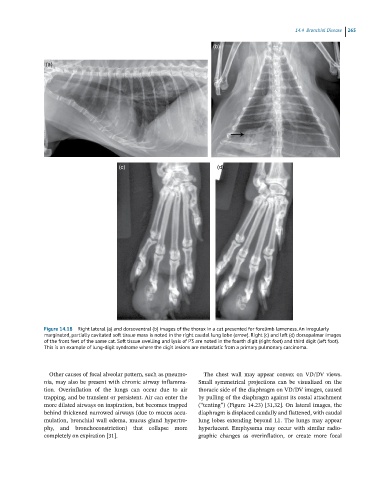
Figure 14.18 Right lateral (a) and dorsoventral (b) images of the thorax in a cat presented for forelimb lameness. An irregularly
marginated, partially cavitated soft tissue mass is noted in the right caudal lung lobe (arrow). Right (c) and left (d) dorsopalmar images
of the front feet of the same cat. Soft tissue swelling and lysis of P3 are noted in the fourth digit (right foot) and third digit (left foot).
This is an example of lung-digit syndrome where the digit lesions are metastatic from a primary pulmonary carcinoma.
Other causes of focal alveolar pattern, such as pneumo- The chest wall may appear convex on VD/DV views.
nia, may also be present with chronic airway inflamma- Small symmetrical projections can be visualized on the
tion. Overinflation of the lungs can occur due to air thoracic side of the diaphragm on VD/DV images, caused
trapping, and be transient or persistent. Air can enter the by pulling of the diaphragm against its costal attachment
more dilated airways on inspiration, but becomes trapped (“tenting”) (Figure 14.23) [31,32]. On lateral images, the
behind thickened narrowed airways (due to mucus accu- diaphragm is displaced caudally and flattened, with caudal
mulation, bronchial wall edema, mucus gland hypertro- lung lobes extending beyond L1. The lungs may appear
phy, and bronchoconstriction) that collapse more hyperlucent. Emphysema may occur with similar radio-
completely on expiration [31]. graphic changes as overinflation, or create more focal