Page 264 - Feline diagnostic imaging
P. 264
268  14  Feline Pulmonary Disease

                                                                     (b)


            (a)


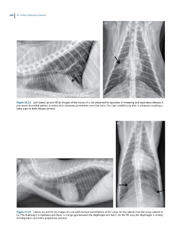
            Figure 14.22  Left lateral (a) and VD (b) images of the thorax of a cat presented for episodes of wheezing and respiratory distress. A
            prominent bronchial pattern is noted, with thickened, prominent bronchial walls. The right middle lung lobe is collapsed, creating a
            lobar sign on both images (arrows).


                                                                             (b)


            (a)


            Figure 14.23  Lateral (a) and VD (b) images of a cat with marked overinflation of the lungs. On the lateral view, the lungs extend to
            L2. The diaphragm is flattened, and there is a large gap between the diaphragm and heart. On the VD view, the diaphragm is tented,
            forming small symmetric projections (arrows).

