
Page 268 - Feline diagnostic imaging
P. 268
272 14 Feline Pulmonary Disease
(b)
(a)
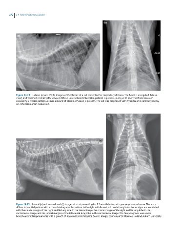
Figure 14.28 Lateral (a) and DV (b) images of the thorax of a cat presented for respiratory distress. The heart is elongated (lateral
view), and widened cranially (DV view). A diffuse, unstructured interstitial pattern is present, along with poorly defined areas of
coalescing alveolar pattern. A small amount of pleural effusion is present. The cat was diagnosed with hypertrophic cardiomyopathy
on echocardiogram evaluation.
(b)
(a)
Figure 14.29 Lateral (a) and ventrodorsal (b) images of a cat presenting for 1.5-month history of upper respiratory disease. There is a
diffuse interstitial pattern with a consolidating alveolar pattern in the right middle and left caudal lung lobes. Lobar signs are associated
with the caudal margin of the right middle lung lobe in the lateral image, the cranial margin of the right middle lung lobe in the
ventrodorsal image, and the lateral margins of the left caudal lung lobe in the ventrodorsal image. The final diagnosis was severe
bronchointerstitial pneumonia with a growth of Bordetella bronchiseptica. Source: Images courtesy of Dr Merrilee Holland, Auburn University.