Page 272 - Feline diagnostic imaging
P. 272
276  14  Feline Pulmonary Disease
            result  in  clinical  infection,  with  severe  bronchitis  or   14.6.4  Feline Infectious Peritonitis
              bronchopneumonia. Radiographic changes of mixed pat-  Feline  infectious  peritonitis  (FIP)  is  characterized  by
            terns  of  diffuse  peribronchial,  interstitial,  and  alveolar
            opacities can be seen [57,71,73].                 fibrinous and granulomatous serositis, protein‐rich serous
                                                                effusions (effusive form), and/or pyogranulomatous lesions
              Pneumonia is the most important clinical manifestation
            of feline toxoplasmosis (Toxoplasma gondii) [74]. Cats are   in  several  organs  (noneffusive  form)  [76,77].  Pleural
                                                                effusion is the most common thoracic manifestation, but a
            the  key  animal  species  in  the  life  cycle  of  this  parasite,
            as  they  are  the  hosts  that  can  excrete  the  oocysts.   pyogranulomatous disease process involving the lungs can
                                                              occur. About 10% of cats with the noneffusive form will
            Immunosuppressed cats may develop clinical disease [75].
                                                              have  thoracic  involvement,  with  localized  pulmonary

                                                                         (b)


               (a)


                        (c)                                  (d)


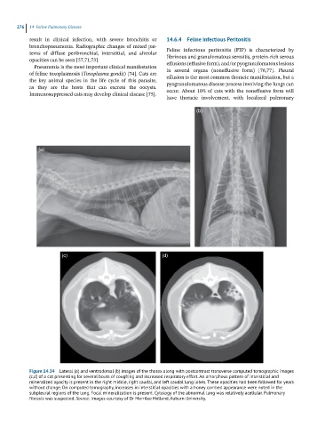
            Figure 14.34  Lateral (a) and ventrodorsal (b) images of the thorax along with postcontrast transverse computed tomographic images
            (c,d) of a cat presenting for several bouts of coughing and increased respiratory effort. An amorphous pattern of interstitial and
            mineralized opacity is present in the right middle, right caudal, and left caudal lung lobes. These opacities had been followed for years
            without change. On computed tomography, increases in interstitial opacities with a honey combed appearance were noted in the
            subpleural regions of the lung. Focal mineralization is present. Cytology of the abnormal lung was relatively acellular. Pulmonary
            fibrosis was suspected. Source: Images courtesy of Dr Merrilee Holland, Auburn University.

