
Page 271 - Feline diagnostic imaging
P. 271
14.6 Pneumonia 275
(b)
(a)
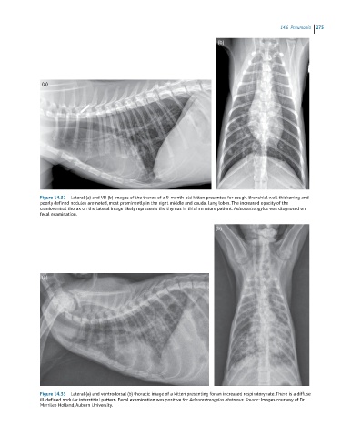
Figure 14.32 Lateral (a) and VD (b) images of the thorax of a 9-month-old kitten presented for cough. Bronchial wall thickening and
poorly defined nodules are noted, most prominently in the right middle and caudal lung lobes. The increased opacity of the
cranioventral thorax on the lateral image likely represents the thymus in this immature patient. Aeleurostrongylus was diagnosed on
fecal examination.
(b)
(a)
Figure 14.33 Lateral (a) and ventrodorsal (b) thoracic image of a kitten presenting for an increased respiratory rate. There is a diffuse
ill-defined nodular interstitial pattern. Fecal examination was positive for Aeleurostrongylus abstrusus. Source: Images courtesy of Dr
Merrilee Holland, Auburn University.