
Page 263 - Feline diagnostic imaging
P. 263
14.4 Bronchial Disease 267
(a)
(b)
(c)
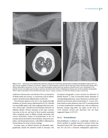
Figure 14.21 Lateral (a) and ventrodorsal (b) thoracic images and transverse postcontrast computed tomography image (c) of a cat
with chronic respiratory disease. On thoracic images, the bronchi appear widened with thickening of the bronchial walls along with a
diffuse interstitial lung pattern. On the computed tomographic image, there are moderate dilated bronchi with thickening of the
smaller airways and patchy ground-glass opacities consistent with chronic inflammatory disease such as feline asthma. Eosinophilic
inflammation was found on the transtracheal wash. Source: Images courtesy of Dr Merrilee Holland, Auburn University.
additional inflammation and infection from accumulation Computed tomography is more sensitive for detection of
of fluids within the airways. A continuous cycle of inflam- dilated bronchi. A ratio of bronchial lumen diameter to
mation/ bronchiectasis/inflammation results [34]. corresponding pulmonary artery diameter in normal anes-
Bronchiectasis appears to be rare in cats, despite the high thetized cats has been determined using CT. A mean bron-
incidence of chronic airway inflammation [23,35]. Affected chial lumen to artery diameter ratio of 0.71 was determined
cats are middle‐aged or old, with coughing as the most com- in normal anesthetized ventilated cats, with no difference
mon clinical sign. Cylindrical bronchiectasis (bronchi are between varying lung lobes. An upper cut‐off value of
diffusely dilated, without tapering) is the most common >0.91 between normal and abnormal cats has been
type seen in the cat, and may be focal (usually associated suggested (Figure 14.27) [37].
with a neoplastic mass) or diffuse (usually associated with
chronic bronchitis). Causes of bronchiectasis in the cat 14.4.2 Broncholithiasis
include chronic bronchitis or bronchiolitis, obstructive neo-
plasia, and bronchopneumonia, with diffuse inflammatory Broncholithiasis is defined as a pathologic condition in
airway disease the most common etiology. which calcified or ossified material is present within the
In some cases, no dilated bronchi are identified radio- bronchial lumen [38]. This condition is rarely reported in
graphically, despite the presence noted at necropsy [36]. cats, but can have a dramatic radiographic appearance.