
Page 258 - Feline diagnostic imaging
P. 258
262 14 Feline Pulmonary Disease
(b)
(a)
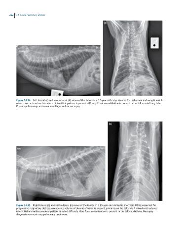
Figure 14.14 Left lateral (a) and ventrodorsal (b) views of the thorax in a 12-year-old cat presented for tachypnea and weight loss. A
mixed unstructured and structured interstitial pattern is present diffusely. Focal consolidation is present in the left cranial lung lobe.
Primary pulmonary carcinoma was diagnosed on necropsy.
(b)
(a)
Figure 14.15 Right lateral (a) and ventrodorsal (b) views of the thorax in a 13-year-old domestic shorthair (DSH) presented for
progressive respiratory distress. A moderate volume of pleural effusion is present, primarily on the left side. A mixed unstructured
interstitial and miliary nodular pattern is noted diffusely. More focal consolidation is present in the left caudal lobe. Necropsy
diagnosis was a primary pulmonary carcinoma.