
Page 255 - Feline diagnostic imaging
P. 255
14.3 Pulmonary eoplasia 259
(b)
(a)
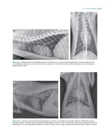
Figure 14.8 Left lateral (a) and ventrodorsal (b) views of the thorax in a 10-year-old cat presented for vomiting and blood in the
stool. Multiple relatively well-circumscribed soft tissue nodules are identified in most lung lobes. Metastatic colon carcinoma was
diagnosed at necropsy.
(b)
(a)
Figure 14.9 Lateral (a) and ventrodorsal (b) radiographs of a female cat presented for respiratory distress. A diffuse hazy nodular
interstitial pattern is present. A pyogranulomatous inflammation was found on the transtracheal wash and was suspected to be due to
Blastomycosis. The cat responded to treatment for fungal infection. Source: Images courtesy of Dr Merrilee Holland, Auburn University.