
Page 252 - Feline diagnostic imaging
P. 252
256 14 Feline Pulmonary Disease
(a) (b)
(c)
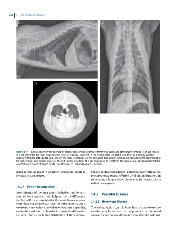
Figure 14.4 Lateral (a) and ventrodorsal (b) radiographs and postcontrast transverse computed tomographic image (c) of the thorax
of a cat presented for fever and anorexia. Alveolar opacity is present in the right middle lung lobe and there is a patchy alveolar
opacity within the left caudal lung lobe on the thoracic images. On the computed tomographic image, air bronchograms are present in
the right middle and caudal aspect of the left cranial lung lobe. This cat responded to treatment for pneumonia cultured as Bordetella
bronchiseptica. Source: Images courtesy of Dr Merrilee Holland, Auburn University.
septal defect) can result in prominent vessels that create an opacity, cardiac size, adjacent abnormalities (rib fractures,
increase in lung opacity. pneumothorax, pleural effusion), will add information. In
some cases, a lung aspirate/biopsy may be necessary for a
definitive diagnosis.
14.1.7 Pattern Determination
Determination of the lung pattern (whether traditional or 14.2 Vascular Disease
nontraditional approach) will help narrow the differential
list, but will not always identify the exact disease process. 14.2.1 Heartworm Disease
More than one disease can have the same pattern, and a
disease process can have more than one pattern, depending The radiographic signs of feline heartworm disease are
on duration and severity. In order to narrow the differential variable, may be transient, or not present at all. Reported
list, other factors, including distribution of the abnormal changes include focal or diffuse bronchointerstitial patterns,