
Page 253 - Feline diagnostic imaging
P. 253
14.3 Pulmonary eoplasia 257
focal or multifocal alveolar opacities, and enlarged periph-
eral pulmonary arteries (Figure 14.12). Using dorsoventral
(DV) or ventrodorsal (VD) images, caudal lobar pulmonary
arteries are considered enlarged if their diameter, meas-
ured at the ninth intercostal space, is greater than 1.6 times
the diameter of the ninth rib [1]. Heartworm‐associated
respiratory disease (HARD) can occur earlier in the disease
process, typically about three months post infection.
Radiographic findings include diffuse or focal bronchoint-
erstitial opacities, with or without peripheral pulmonary
arterial enlargement. Pure bronchial and pure interstitial
infiltrative patterns are less common, and alveolar patterns
are uncommon. Thoracic radiographs may be completely
normal [2–6].
14.3 Pulmonary Neoplasia
14.3.1 Primary Pulmonary Neoplasia
The radiographic appearance of primary pulmonary neo-
plasia is extremely variable, likely more so than in the
dog (Figures 14.13–14.15) [7–13]. The most common pri-
mary pulmonary tumor in the cat is adenocarcinoma,
with squamous cell carcinoma and bronchoalveolar car-
cinoma noted less commonly [14]. These tumors cannot
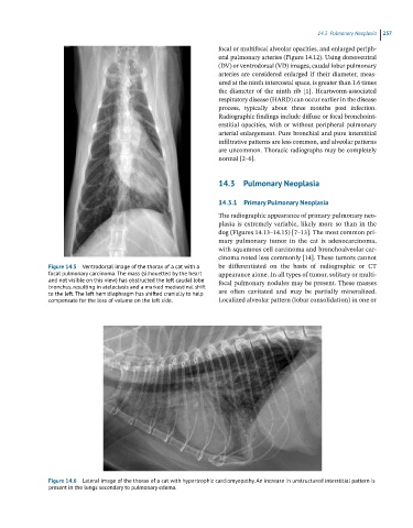
Figure 14.5 Ventrodorsal image of the thorax of a cat with a be differentiated on the basis of radiographic or CT
focal pulmonary carcinoma. The mass (silhouetted by the heart appearance alone. In all types of tumor, solitary or multi-
and not visible on this view) has obstructed the left caudal lobe focal pulmonary nodules may be present. These masses
bronchus, resulting in atelectasis and a marked mediastinal shift
to the left. The left hemidiaphragm has shifted cranially to help are often cavitated and may be partially mineralized.
compensate for the loss of volume on the left side. Localized alveolar pattern (lobar consolidation) in one or
Figure 14.6 Lateral image of the thorax of a cat with hypertrophic cardiomyopathy. An increase in unstructured interstitial pattern is
present in the lungs secondary to pulmonary edema.