
Page 250 - Feline diagnostic imaging
P. 250
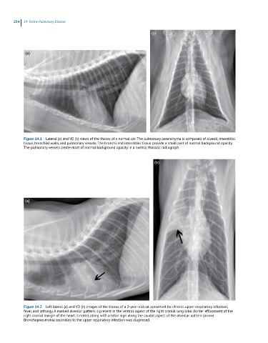
254  14  Feline Pulmonary Disease

                                                                      (b)


             (a)


            Figure 14.1  Lateral (a) and VD (b) views of the thorax of a normal cat. The pulmonary parenchyma is composed of alveoli, interstitial
            tissue, bronchial walls, and pulmonary vessels. The bronchi and interstitial tissue provide a small part of normal background opacity.
            The pulmonary vessels create most of normal background opacity in a normal thoracic radiograph.


                                                                        (b)


            (a)


            Figure 14.2  Left lateral (a) and VD (b) images of the thorax of a 2-year-old cat presented for chronic upper respiratory infection,
            fever, and lethargy. A marked alveolar pattern is present in the ventral aspect of the right cranial lung lobe. Border effacement of the
            right cranial margin of the heart is noted, along with a lobar sign along the caudal aspect of the alveolar pattern (arrow).
            Bronchopneumonia secondary to the upper respiratory infection was diagnosed.